Brian Choi, Bio-application scientist
For more information, please contact app@parksystems.com
Cell has different mechanical property depending on the physiological processes (cell differentiation, growth, adhesion, etc.) and under various pathogenesis (oxidative stress, virus infection, parasites, etc.). Force distance spectroscopy of atomic force microscope (AFM) allows the cell’s mechanical property analysis on the surface at nano-level, and subsurface layers of cells by probing the cell membrane with high resolution cantilever tip (<10 nm). Recording the force value and vertical deflection of the cantilever, the force value versus distance graph (force-distance curve) can be acquired between the probe and the surface. Analyzing the force-distance curve with the Hertz model, the cell’s mechanical property can be calculated quantitatively.
- Analyze the cell’s mechanical property on the surface at nano-level with force-distance spectroscopy of atomic force microscope (AFM)
- Acquire the quantitative mechanical property of cell, by calculating Young’s modulus of cell with the Hertz model
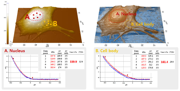
Single Cell (C2C12) Young’s Modulus Measurement: Nucleus vs. Cell body

In this study, the quantitative mechanical properties at two different area (nucleus and cell body) of C2C12 cell were acquired by applying the acquired five force-distance curves from each area into the Hertz model. The high resolution AFM imaging of cell allows the determination of the probing position of cantilever tip accurately. The Young’s modulus of cell was determined at the point where the cantilever deflection reached 3.5 nN in each force-distance curve. In nucleus, the Young’s modulus was calculated to be 339.9 kPa (± 32.9) while the result from the cell body was 161.4 kPa (±29.0). It is obvious that at the nucleus area, the mechanical property of cell is about two times bigger or to say that it’s two times stiffer than at the cell body.
Park Cell Analysis Systems

|

|

|
|
| Park NX12-Bio | Park NX10 | Park XE7 | |
| Scanning Ion Conductance Microscopy (SICM) | |||
| Atomic Force Microscopy (AFM) with liquid probe hand | |||
| Inverted Optical Microscopy (IOM) | |||
| Live Cell Chamber |

