Brian Choi, Bio-application scientist
For more information, please contact app@parksystems.com
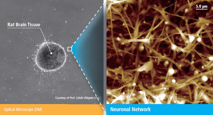
Sample courtesy : Prof. Ushiki, Niigata Univ.
Scanning ion conductance microscopy (SICM) is a new nanoscalemicroscopy specially developed for biological object observation in liquid environments. The nanoscale high-resolution of SICM enables visualizing the details of biologically important features of a size not observable with conventional optical microscopy. SICM’s in-liquid imaging capability allows biologists to observe morphology of biological features as never before possible with scanning electron microscopy (SEM). SICM opens up a new era of microscopic research for biology and life science.
- Discover the physiological phenomena of cells at nanoscale
- Directly acquire a cell’s morphology in-buffer without sample preparation
- Image delicate cell morphology from sub-cellular to tissue level

Scanning Ion Conductance Microscope
Neuronal Netwrok, measured in fluid

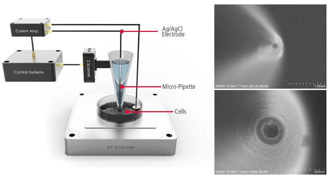
Configuration of SICMNano-pipette End Opening (SEM image)
SICM is a type of scanning probe microscopy and was invented in 1989 in Prof. Hansma’s group. Unlike optical microscopes, SICM uses a glass nanopipette as a sensitive probe with an electrode inside of it. The glass pipette detects nearby surfaces via a decrease in the ion current flow through the pipette.
Park Cell Analysis Systems

|

|

|
|
| Park NX12-Bio | Park NX10 | Park XE7 | |
| Scanning Ion Conductance Microscopy (SICM) | |||
| Atomic Force Microscopy (AFM) with liquid probe hand | |||
| Inverted Optical Microscopy (IOM) | |||
| Live Cell Chamber |

