Brian Choi, Bio-application scientist
For more information, please contact app@parksystems.com
Sample courtesy: Dr. Muhanna, King Abdulaziz City for Science and Technology.
Scanning ion conductance microscopy (SICM) is a practical cell research tool that has an exclusive in-liquid cell imaging capability. In a liquid environment, SICM can directly, yet noninvasively, acquire cell surface morphology. The in-liquid imaging capability without physical contact allows using SICM for various cell study topics such as cell division, fusion, and other fundamental physiological phenomena. SICM is especially useful for studying cell-to-cell interaction such as cell adhesion and cell signalling. By acquiring cellular morphology at the connective position between two cells with nanometer spatial resolution, it is possible to study interactive functions and biological pathways of the cell at the molecule level.
- Noninvasive imaging allows monitoring the cell’s real physiology in a live environment
- Observe morphological dynamics of cell-to-cell interaction
- Monitor cell adhesion molecule behavior
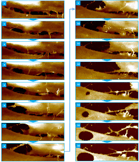
SICM will ultimately help researchers understand the mechanism of cell physiology in real time. One of the distinguishing functions of cell-to-cell interaction is cell adhesion, which not only corresponds to cell development but also to structural maintenance, cell migration, and pathogenesis of infectious organisms. Such adhesion molecule morphology of cells in live environments could not have been captured previously due to limitations of optical microscopes. SICM’s live cell surface imaging allows examining accurate morphological characteristics of adhesion molecules, whose physiological behavior is only detectable in living cells. In addition, time-lapse SICM imaging can be a great help in understanding the behavioral mechanisms of adhesion molecules at a fundamental level.
 Live Whole Cell Imaging with SICM
 Cell Adhesion Molecule Imaging in Living Cell to Cell Interaction
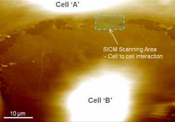
In this study, a cultured brain tumor cell’s adhesion molecule was visualized by SICM to investigate cell-to-cell interactional behavior. At first, the edge position of the brain tumor cell whose membrane is overlapped with the end of the other cell was observed successfully. In the acquired image, connective projections between cell A and B were identified (in green). These projections were observed to hold other cell membranes and ranged in width from 100nm to 300nm.
Park Cell Analysis Systems

|

|

|
|
| Park NX12-Bio | Park NX10 | Park XE7 | |
| Scanning Ion Conductance Microscopy (SICM) | |||
| Atomic Force Microscopy (AFM) with liquid probe hand | |||
| Inverted Optical Microscopy (IOM) | |||
| Live Cell Chamber |

