Brian Choi, Bio-application scientist
For more information, please contact app@parksystems.com
Data Reference: Gordon Jung, Application Scientist
Many toxins can cause acute epithelial inflammation and toxicity. However, the mechanism of such acute toxicity is difficult to elucidate and prove by experiment. Observing the negative effects of such toxicity at the single-cell level is a great help in explaining the mechanism in an intuitive manner. The accurate living cell membrane imaging of scanning ion conductance microscopy (SICM) can detect and visualize the negative effects in morphology occurring as a result of toxins.
- Accurate living cell membrane morphology imaging for visualizing single-cell cytotoxicity
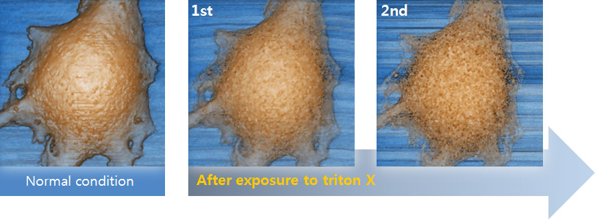
- Cell membrane damage by exposure to Triton X is clearly monitored over time, which is not detectable in optical microscopic view
Cell Membrane Imaging After Detergent Exposure

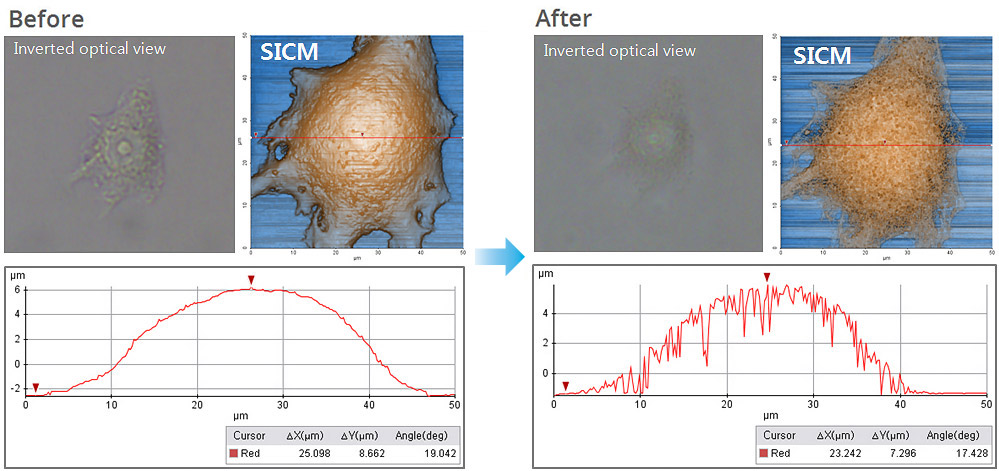
In this experiment, the early response of a living epithelial cell after exposure to Triton X (a nonionic detergent having highly toxic effects on cells) was investigated by SICM. Continuous SICM imaging revealed that cell membrane deformation occurred after Triton X exposure, and it showed the increased number and size of holes on the cell membrane.
Morphology Analysis in Line Data

Cell membrane damage caused by Triton X is more distinctively identified by comparing the morphology analysis in single-line data (the damage is not distinguishable in inverted optical microscopic view). The line data analysis shows the depth and width of holes in the membrane in micrometers, while no hole structure appears in the line data of the cell image before Triton X exposure.
Park Cell Analysis Systems

|

|

|
|
| Park NX12-Bio | Park NX10 | Park XE7 | |
| Scanning Ion Conductance Microscopy (SICM) | |||
| Atomic Force Microscopy (AFM) with liquid probe hand | |||
| Inverted Optical Microscopy (IOM) | |||
| Live Cell Chamber |

