Featured Application
Non-invasive In-liquid Imaging by Ion Conductance Microscopy (ICM)
With the existing research tools available in the market today, it has been virtually impossible to perform nanoscale imaging of single live cells. In particular, cellular membrane surfaces are transparent to optical microscopy methods, and too soft to produce any contrast or even endure probing by AFM method. Ion Conductance Microscopy (ICM) is the only non-invasive in-liquid scanning probe technique that does not apply any force over its sample surface, making it ideal for nanoscale imaging of single live cells, in particular soft cellular membranes. It can be further adapted to enable a host of powerful applications in nano-manipulated electrophysiology and cell motility studies, whose examples include targeted localized stimulation, monitoring, and drug delivery at the cellular level. Click here for further information on the mechanism and applications of ICM.
Featured Product
XE-Bio™: Cell Discovery Like Never Before
XE-Bio™provides the most detailed and integrated information on nanoscale biological surfaces available today, enabling breakthrough research on single live cells by connecting their detailed structural features to cellular functions. Distinguished researchers from Harvard Medical School, Johns Hopkins University, NIH and other major universities and research institutes expressed interest in the versatile use of ICM;ICM is the only non-invasive in-liquid scanning probe technique that does not apply any force over its sample surface, making it ideal for nanoscale imaging of soft cellular membranes. The ICM also enables localized functional studies on single live cells, including functional mapping on membrane features. Click here for the detailed product information.
News and Events
XE-Bio™ Brings Promise in the Fight against Hepatitis C.
Suwon, South Korea, Feb 12, 2010 - The fight against the liver disease hepatitis C has been something of an impasse for years, with more than 150 million people currently infected, and traditional antiviral treatments causing nasty side effects and often falling short ofa cure. Park Systems’ XE-Bio™helped researchers at Stanford University discover a vulnerable step in the virus’ reproduction process that in lab testing could be effectively targeted with an obsolete antihistamine (Click here for the latest news of vaccine development for hepatitis C). XE-Bio™ successfully imaged and analyzed two of the compounds that most potently inhibit vesicle aggregation revealed their mechanism of action on 4BAH2 function. These results provide new insight into the molecular mechanism of HCV replication platform assembly, and demonstrate the utility of a novel small-molecule anti-HCV strategy.Click here for the rest of the news article.
XE-3DM™ Made the Cover of Microscopy and Analysis Magazine.
Suwon, South Korea, Feb 11, 2010 - Park Systems’ XE-3DM™ was featured on the front cover of Microscopy and Analysis (M&A) magazine. The 3D image of an overhang structure on the front cover was imaged with Park Systems’ new 3D atomic force microscope. The new XE-3DM™ is designed for overhang and trench profiles, sidewall roughness imaging, and critical angle measurements. Click here for the rest of the news article.
Upcoming Exhibitions
2010 Biophysical Society Meeting, February 20th ~ 23rd, San Francisco, USA
Park Systems is a proud official sponsor of opening reception at the 2010 Biophysical Society Meeting in San Francisco from February 20th to 23rd. Come and visit us at the Booth 525. See the XE-Bio™, the latest innovation in live cell imaging and learn about non-invasive, high resolution, in-liquid imaging technique for live cells.
APS Meeting, March 15th ~ 19th, Portland, USA
We cordially invite you to visit our booth (#210) at the 2010 APS March Meeting in Portland, USA from Mar. 15th-19th, 2010. See the award winning research-grade XE-100, and learn about artifact free imaging by Crosstalk Elimination (XE) and ultimate AFM resolution by True Non-Contact mode. The XE-100 is our flagship AFM with reduced drift rate and motorized sample stage for the Step-and-Scan Automation.
ACS Spring Meeting, March 21st ~ 25th, San Francisco, USA
Come and visit our booth (#428) at the 2010 ACS Spring Meeting in San Francisco, USA from Mar. 21st-25th, 2010. See the new XE-100, research-grade AFM, with reduced drift rate and motorized sample stage with Step-and-Scan Automation that provides the ultimate AFM/SPM performance in Non-Contact nanoscale metrology.
Images from Crosstalk Eliminated (XE) AFM
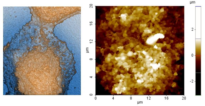
The image of a cancer cell of lung by XE-Bio™
The surface of a live lung cancer cell that imaged in buffer solution using ion conductance microscope shows the rough surfacestructure typical of cancer cells. A traditional AFM is not capable of imaging such membrane structure (without fixation) due to the large interaction force between the tip and the sample.
For further inquiries please contact us via info@parkafm.com
|


