Make the most out of your time: the benefits of easy automation for AFM

Make the most out of your time: the benefits of easy automation for AFM (StepScanTM, AdaptiveScanTM, FastApproachTM)
Wednesday, 2 June, 2021
- 10:00 am – 11:30 am
(GMT)
London, Dublin - 11:00 am – 12:30 pm
(CET)
Berlin, Paris, Rome - 18:00pm – 19:30 pm
[UTC+9]
Seoul, Tokyo

Abstract:
Atomic Force Microscopy is well known for acquiring images with resolution better than diffraction-limited light microscopies in practically any environment. Versatility of AFM based methods makes it an indispensable tool for researchers in numerous areas such as nanometrology, material science, and biology. When using an AFM, researchers spend significant time on tedious tasks such as aligning the optical beam onto cantilever, finding an area of interest on the sample, and optimizing imaging parameters.
Time consuming trial-and-error optimization of the generic height imaging parameters such as scan rate, gain settings, and setpoint is always required to get some reliable AFM data. Advanced AFM operation modes involve control of additional variables. Interdependencies of the parameters make it challenging to find optimum operating conditions in the parameter space. Moreover, a single instrument is often shared among several researchers of varying AFM skills. Throughput of AFM data by a researcher is further reduced either due to limited software tools or a steep learning curve of the user interface.
Park Systems’ SmartScan software provides parameter automation while also allowing full manual control in a user-friendly interface. This makes Park AFMs efficient tools for novices and experts alike. Unique automation features such as FastApproachTM, AdaptiveScanTM, and StepScanTM allow users to make the most out of their AFM time. In this webinar we will demonstrate the use of these automation features.
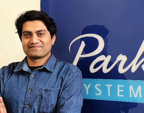
Presented By :
Abdul Rauf, Application Scientist at Park Systems Europe
Abdul is an Application Scientist at Park Systems Europe, where he supports development of AFM solutions for customers. He had his training as a polymer materials’ engineer with emphasis on elastomer blends and composites. Abdul has expertise in characterization of macromolecular systems at interfaces. He worked on his doctoral thesis in the group of Prof. Jürgen P. Rabe in Humboldt-Universität zu Berlin, where he acquired expertise in morphological and nanomechanical characterization of thin films confined in interfaces. His work in Berlin also included study of two-dimensional materials such as monolayers of Graphene, Hexagonal Boron Nitride, and Transition Metal Dichalcogenides as sensors for strain transfer across atomic interfaces.
パーク・システムズ#9 研究⽤AFM: AFMプローブの選択方法
2021 Park AFM Certification Course: Lecture #4: Nanoindentation
Make the most out of your potential: the benefits of single-pass sideband KPFM

Make the most out of your potential: the benefits of single-pass sideband KPFM
Wednesday, 23 June, 2021
- 10:00 am – 11:30 am
(GMT)
London, Dublin - 11:00 am – 12:30 pm
(CET)
Berlin, Paris, Rome - 17:00 am – 18:30 pm
[GMT+8]
Beijing, Singapore - 18:00pm – 19:30 pm
[UTC+9]
Seoul, Tokyo
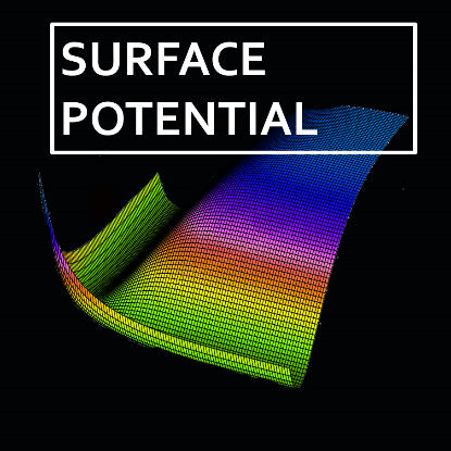
Abstract:
Atomic Force Microscopy (AFM) has become an essential tool for research in a large range of fields of sciences. AFM provides higher resolution when compared to other microscopy techniques. Its applicability on the most diverse samples makes it the ideal instrument to explore properties of materials at their interfaces, to study biological material and processes, and to understand fundamental properties of condensed matter.
Due to its high performance in terms of resolution, AFM is largely used to perform structural and topographical characterization. Nevertheless, there is much more than just topography under an AFM tip. Once brought close to the sample, the AFM probe is sensitive to a series of interactions which depend on local properties of the material under inspection. It is possible to prepare AFM experiments in such a way that quantitative data about these properties can be collected, empowering the researcher with a set of new and exciting information to link with topography.
Among the advanced modes AFM is capable of, Kelvin Probe Force Microscopy (KPFM) allows retrieving data on the electrical potential at the sample surface. KPFM is then the ideal local probe technique to correlate structural features and material composition of the observed sample with the variation of such potentials, with possible applications in many different fields like nano- and optoelectronics, metal corrosion and energy storage.
Park Systems has implemented single-pass sideband KPFM by default in all research tools of its NX series. This advanced mode enables users to perform highly precise, quantitative mapping of the potential distribution at the same time than topography.
In this course, we will show details and advantages of the KPFM technique, together with a demonstration of its ease of use.
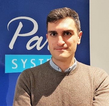
Presented By :
Dr. Andrea Cerreta, Application Scientist Park Systems Europe, Mannheim, Germany
acerreta@parksystems.com
Dr. Andrea Cerreta is an Application Scientists at Park Systems Europe, where he focuses on application development and support for the academic sector. He received his Ph.D. in Physics from the Ecole Polytechnique Fédérale de Lausanne (EPFL), Switzerland. He did his further doctoral work at the Solid State Physics Group of Université de Fribourg, Switzerland, which focused on studying electrical and magnetic properties of organic spin valves and spin polarized currents in superconducting materials, grown by means of Pulsed Laser Deposition, and characterizing the DC and AC transport properties of magnetic and superconducting samples. His expertise also spans the Frequency Modulation Atomic Force Microscopy in UHV for the study of biomolecules.
