Microscopía de fuerza de sonda Kelvin de banda lateral para caracterización avanzada de materiales (Spanish/español)
Microscopía de fuerza de sonda Kelvin de banda lateral para caracterización avanzada de materiales.
Viernes, Apr 30, 2021
*Note: This webinar will be presented in Spanish/español
- 10:00 AM
(CDT)
MEXICO / COLOMBIA - 8:00 AM
(PST)
LOS ANGELES / TIJUANA - 13:00 PM
(UCT)
BRASIL / ARGENTINA - 5:00 PM
(CEST)
ESPANA
La microscopía de fuerza de sonda Kelvin de banda lateral (KPFM) utiliza la intermodulación de una fuerza impulsora electrostática y una fuerza impulsora mecánica para convertir la frecuencia electrostática a la primera resonancia de flexión, donde el factor de alta calidad de la resonancia produce una medición más sensible. La señal KPFM de banda lateral se calcula utilizando una interacción local entre el ápice de la punta y la muestra en lugar de una interacción total entre el voladizo y la muestra, mejorando la resolución espacial sobre otras variaciones de la técnica. Únase a nosotros como ingeniero de servicios técnicos en Park Systems que cubre los conceptos básicos y más detalles de KPFM de banda lateral, incluidas las compensaciones y sugerencias de imágenes para Park AFM con varias imágenes de materiales avanzados.
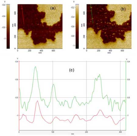
Figura: Diferencia de potencial de superficie usando KPFM de banda lateral en alcano semifluorado usando (a) un voladizo más corto y duro y (b) un voladizo más largo y suave. Perfiles de línea (c) que muestran una resolución más alta con un voladizo más largo y suave (b) (Referencia: Imágenes de potencial de superficie a través de microscopía de fuerza de sonda Kelvin de banda lateral (Parksystems.com)

ACERCA DE NUESTRO PANELISTA :
Armando Melgarejo es ingeniero de soporte en Park Systems
Armando Melgarejo es ingeniero de soporte en Park Systems, donde se dedica a la instalación y atención de AFM al nivel de investigación y posgrado. Tiene un título en ingeniería en Biotecnología de la Universidad Autónoma de Querétaro, México. Durante sus estudios hizo un semestre de investigación en la Universidad Tecnica de Czechia en Praga, Republica Checa; donde se especializo en nanotecnología y biología molecular. Entre sus áreas de experiencia se encuentran diversas técnicas de caracterización (RAMAN, AFM and IR), inmovilización de enzimas, genética y biología celular.
Nanoscale Electrochemistry Study using Scanning Electrochemical Cell Microscopy (SECCM)
Nanoscale Electrochemistry Study using Scanning Electrochemical Cell Microscopy (SECCM)
Thesday, March 9, 2021
- 10:30 am
(PKT)
Islamabad, Pakistan - 11:00 am
(IST)
Bangalore, India - 13:30 pm
(SGT)
Singapore, Singapore - 16:30 pm
(ADET)
Sydney, Australia
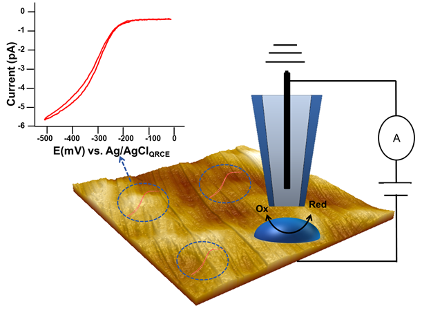
Linear cyclic voltammograms obtained from dispensing droplets of Ru(NH3)6Cl3 solution at a HOPG surface in the SECCM format with a glass pipette using a Park NX12 system . The CV was recorded at a sweep rate of 10 mV/s.
The application staff of Park Systems will present an introduction to Scanning electrochemical cell microscopy (SECCM). SECCM is a nano-electrochemical scanning probe technique that creates a confined electrochemical cell (droplet) via meniscus contacting the surface by employing an electrolyte filled micropipette containing a quasi-reference counter electrode (QRCE). It allows researchers to investigate the local electrochemical properties of the electrode surfaces by directly mapping the electroactivity with nanoscale resolution. The electrochemically reversible [Ru (NH3)6]3+/2+ electron transfer process at a highly ordered pyrolytic graphite (HOPG) surface was recorded using the Park NX12 AFM system in this study.
Join us as the technical services engineer at Park Systems, explain the basics of SECCM and quantitative electroanalysis at the nanoscale using the Park NX12 AFM System.
Presented By :
Jiali Zhang, Ph.D., Applications Engineer, Park Systems
Jiali Zhang, Ph.D., is an engineer for Park Systems, where she focuses on the installation and support of AFM systems for Park’s research user base. She is also responsible for researching and writing technical papers and application notes for publication and presentation at scientific conferences. She received her Ph.D. in Analytical Chemistry from the University of California, Davis, and holds a B.S. in Applied Chemistry from Donghua University in Shanghai, China. Her expertise spans numerous microscopy techniques, and areas of study have also included biological systems and 3D printing technologies.
PinPointing peptides: Unveiling molecular structure and nanomechanical properties of peptide nanotubes
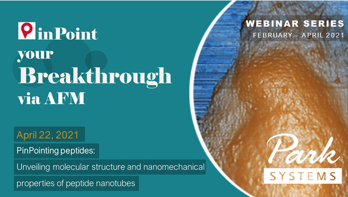
PinPointing peptides: Unveiling molecular structure and nanomechanical properties of peptide nanotubes
Thursday, 22 April, 2021
- 10:00 am – 11:30 am
(GMT)
London, Dublin - 11:00 am – 12:30 pm
(CET)
Berlin, Paris, Rome - 18:00pm – 19:30 pm
[UTC+9]
Seoul, Tokyo

The self-assembly of short peptides has been recently established as a facile route to various macromolecular nanostructures with promising applications in tissue engineering, biomedical devices and drug delivery. Some of these nanostructures may also be used as degradable scaffolds for the fabrication of metal nanowires. Self-assembled peptides can form an array of nanostructures including nanofibrils, nanotubes, nanospheres and vesicles dependent on the constituent peptide or environmental conditions during assembly. They also display a range of nanomechanical and, as has been recently shown [1], strong piezoelectric properties. At the same time, unveiling their molecular structure and mechanism of peptide self-assembly is crucial in understanding the precise molecular routes for medical conditions associated with protein misfolding, e.g. Alzheimer disease.
Therefore, peptide nanotubes and similar structures are ideal objects for correlative AFM imaging – an approach to combine both molecular resolution and nanomechanical/nanoelectrical data within the same experiment. Such approach could lead to the ultimate goal – correlating molecular structure of a peptide nanotube with its functional properties. Something that has not been achieved so far
This webinar will focus on practical aspects of acquiring high resolution and nanomechanical data with both PinPointTM and True Non-ContactTM modes on a range of peptide nanotubes.
[1] A. Kholkin , N. Amdursky , I. Bdikin , E. Gazit , G. Rosenman , Strong piezoelectricity in bioinspired peptide nanotubes, ACS Nano (2010) 610–614
Presented By :
Dr. Vladimir Korolkov, Senior Application Scientist at Park Systems UK
Vladimir received his PhD in Chemistry from Moscow University in 2008. Then, he moved to the University of Heidelberg and specialized in X-ray photoelectron spectroscopy of thin films, following by the position at the University of Nottingham, where he discovered his passion for Scanning Probe Microscopy (SPM), and became a strong advocate of SPM techniques to unlock structure and properties at nanoscale. He pioneered the use of higher eigenmodes of standard cantilevers to routinely achieve resolution that was previously thought to be exclusively limited to STM and UHV-STM. Vladimir published more than 40 scientific papers, including three in Nature family journals. He left academia in 2018 to contribute to the industrial site of SPM technology.
Surface Potential Imaging via Sideband KPFM
Surface Potential Imaging via Sideband KPFM
Thursday, 23 February, 2021
- 11:00 am
(PKT)
Islamabad, Pakistan - 11:30 am
(IST)
Bangalore, India - 14:00 pm
(SGT)
Singapore, Singapore - 17:00 pm
(AEDT)
Sydney, Australia
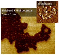
Sideband Kelvin Probe Force Microscopy (KPFM) is a new mode from Park Systems that allows simultaneous imaging of topography and surface potential with a high spatial resolution and improved sensitivity compared to conventional lift mode and other KPFM methods. In this method, the topography is detected at the cantilever's resonance, while the KPFM signals are detected at the sideband’s frequency, a few kHz off from the cantilever's resonant frequency. Therefore, both signals can be measured simultaneously, significantly reducing the acquisition time while improving the surface potential imaging sensitivity.
This webinar will introduce Sideband KPFM, using F14H20 as an object of study for the surface potential measurements.
Presented By :
Armando Melgarejo Technical Support Engineer, Park Systems
Armando Melgarejo is an engineer for Park Systems, where he focuses on the installation and support of AFM systems for Park’s research user base. He holds a B.S. in Biotechnology Engineering from Autonomous University of Queretaro, Mexico. During his studies, he also spent a semester doing research in nanotechnology and molecular biology at Czech Technical University in Prague, Czech Republic. Other areas of expertise include diverse characterization techniques (AFM, SEM, RAMAN and IR), genetics and molecular and cell biology.
Nanoscale exploration of doping patterns in electronic devices (via PinPoint SCM)
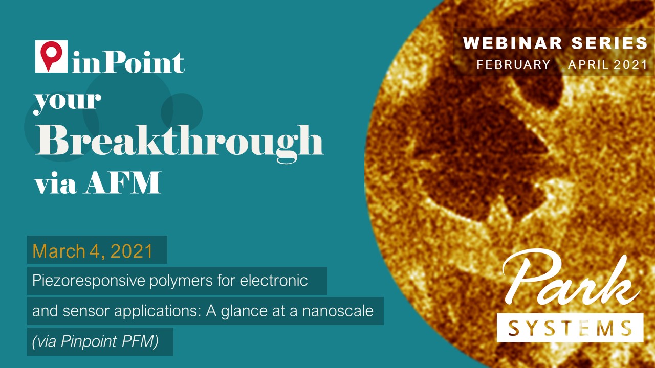
Piezoresponsive polymers for electronic and sensor applications: a glance at a nanoscale (via Pinpoint PFM)
Thursday, 4 March, 2021
- 10:00 am – 11:30 am
(GMT)
London, Dublin - 11:00 am – 12:30 pm
(CET)
Berlin, Paris, Rome - 18:00pm – 19:30 pm
[UTC+9]
Seoul, Tokyo

With the integration of piezoelectric polymers in sensors and electronic devices, such as transducers, an accurate structural and electromechanical characterization down to the nanoscale becomes vital. As real-space, high resolution imaging technique, atomic force microscopy (AFM) accesses not only morphological sample information, but also resolves functional properties including nanomechanics, piezo-/ferroelectricity and electrical potential. Therefore, AFM is ideally suited for a holistic investigation of functional polymer samples with a nanometer resolution. Here, we demonstrate the capabilities of Park Systems’ research AFMs to image the piezoelectric and nanomechanical properties of PVDF fibers simultaneously via PinPointpiezoelectric force microscopy (PFM). PinPoint PFM prevents tip or sample damage via the elimination of shear forces and reduces topography crosstalk due to well defined tip-sample contact. The webinar will include a live measurement on our small sample NX10 research AFM.
Presented By :
Ilka Hermes, Principle Scientist Park Systems Europe, Mannheim, Germany
Ilka is the principle scientist at Park Systems Europe, where she maintains and supports scientific collaborations to establish new research projects. Prior, she worked at the Max Planck Institute for Polymer Research (Main Germany) in the group of Stefan Weber to investigate perovskite solar cells with electrical Atomic Force Microscopy (AFM) modes, and at the Johannes Gutenberg University Mainz in the group of Angelika Kühnle to characterize liquid-solid interfaces with high resolution AFM. Ilka’s primary fields of expertise include Piezoresponse Force Microscopy (PFM), Kelvin Probe Force Microscopy (KFM), and conductive AFM on semiconducting and/or ferroelectric devices.